Chapter 2: Neuroanatomy
2nd edition as of August 2022
Chapter Overview
In the last chapter, we introduced psychoactive drugs, drugs that can influence our behavior and physiological functions. It is important to realize that a drug cannot make the body do something it is incapable of doing. Drugs cannot give you the ability to walk through walls or fly through the air. Instead, they interact with the systems that regulate our body functions, causing us to feel more awake (caffeine), lose our inhibition (alcohol), or experience less pain (opioids).
To understand how drugs work, you must understand how the body normally works. The next few chapters focus on the human nervous system at three different levels: the overall structure; the individual nerve cells; and neurotransmitters. This crash course in neuroscience is simplified and streamlined here. It can be a lot of information to take in, so make sure to give yourself time to process it. Take advantage of the optional videos listed in this module on Canvas to reinforce your learning.
In Chapter 2, we will cover the basic structure of the nervous system and how it is organized. We will also highlight a few regions that are relevant to drug use and the development of drug dependence. This is a short chapter, but a lot of new terminology will be introduced. Make sure you test yourself on the divisions of the nervous system and regions of the brain. This way, you will be able to differentiate between them and define their functions. Later we will see how drugs produce different effects by acting on different molecular targets in different parts of the brain.
Chapter Outline
2.1. Overview of the Nervous System
- 2.1.1. The Structure of the Nervous System
- 2.1.2. The Divisions of the Peripheral Nervous System
- 2.2.1. Cerebral Cortex
- 2.2.2. Thalamus, Hypothalamus, and Limbic System
- 2.2.3. Cerebellum and Brainstem
2.3. Gray Matter vs. White Matter
Chapter Learning Outcomes
- Describe the organization of the nervous system and distinguish nerves and neurons.
- List and describe the structures of the brain.
- Describe the two types of brain tissue.
2.1. Overview of the Nervous System
Section Learning Objectives
- Explain the organization and role of the nervous system.
- Define a nerve and distinguish it from a neuron.
- Distinguish between central and peripheral nervous systems, afferent and efferent nerves, and the somatic and autonomic nervous systems.
- Explain how the sympathetic and parasympathetic nervous systems work.
- Describe dual innervation.
If you’re taking this class, you have likely studied the nervous system before as part of a biology course or introductory psychology course. To refresh, the nervous system is responsible for transmitting signals and coordinating activity between different parts of the body. Understanding how the nervous system functions is important for this class because all the psychoactive drugs we will study in this course will influence the body through the nervous system.
To start, we will provide a basic overview of the nervous system and its major divisions. To prepare for this, please watch this brief video that summarizes the upcoming information in a concise and easy-to-understand format:
Alila Medical Media – Overview of the Nervous System [4:10]
2.1.1. The Structure of the Nervous System
In vertebrates like humans, the nervous system can be divided into two main parts.
The first is the central nervous system (CNS), which consists of the brain and spinal cord. The CNS is “central” because it is responsible for the regulation of bodily functions. It receives signals from different parts of the body, integrates and processes the information, then coordinates a response. By comparison, the peripheral nervous system (PNS) deals with transmission, carrying signals between the CNS and different parts of the body. It is involved in everything outside (the periphery) of the CNS.
The PNS consists of ganglia and nerves, which are bundles of axon fibers that transmit electrical impulses. Peripheral nerves can contain either afferent nerve fibers, efferent nerve fibers, or a mix of afferent and efferent nerve fibers. Afferent nerve fibers (from ad in Latin for “toward” and ferre for “to carry”) convey sensory information from the body to the CNS. Efferent nerve fibers (from ex in Latin for “from” and ferre for “to carry”) convey motor commands from the CNS to various muscles and glands. Mixed nerves contain both afferent and efferent nerve fibers. (A good way to remember the difference is that afferent nerves arrive at the CNS, while efferent nerves exit the CNS.) Each nerve fiber is the axon of a nerve cell or neuron, which we will cover in detail later. Neurons are also present in the CNS, forming a web of connections that can process information and coordinate sophisticated responses. We will explore the CNS in the second half of the chapter. For now, we will discuss the PNS.
2.1.2. The Divisions of the Peripheral Nervous System
The PNS itself can be further separated into two main divisions. The somatic nervous system is associated with voluntary movement. It transmits sensory information from the body (from soma in Greek, hence the name) and conveys motor commands to skeletal or voluntary muscles. Without somatic nerve innervation, the skeletal muscles are incapable of movement, undergo paralysis, and eventually atrophy. The autonomic nervous system, as its name suggests, is associated with automatic or unconscious functions and is always active. It transmits sensory information to smooth muscle, cardiac muscle, and glandular tissues which constitute the internal organs (viscera). This is why the system is sometimes called the visceral nervous system. Unlike skeletal muscles, autonomic tissues are not dependent on the CNS for their function, although they can be regulated by the CNS.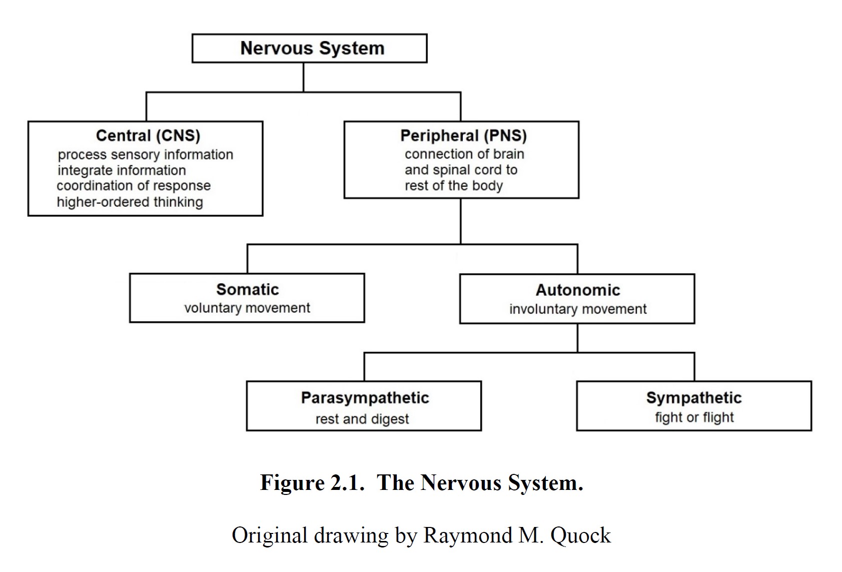
The autonomic nervous system has two branches. The sympathetic nervous system is our “fight-or-flight” system. When we are stressed or perceive danger, this system increases our heart rate and blood pressure, increases blood flow to the skeletal muscles, dilates our pupils, and inhibits digestion. The parasympathetic nervous system is our “rest-and-digest” system. In times of relaxation, it slows our heart rate, constricts our pupils, and stimulates digestion. As you can see, the two branches tend to have opposite or counteracting effects, working together to adapt your body to different situations. The sympathetic nervous system allows for quick mobilization and response, while the parasympathetic nervous system brings the body back to its default state.
Why is it called the “sympathetic” nervous system?
Although we usually use the word sympathy to mean feeling pity or sharing feelings with someone else, the sympathetic nervous system is not related to this concept. Instead, the word sympathetic was used in the past because it was noted that organs were responding in the same way to the same thing as if they were working together. It is from this less common definition of the word sympathy (“feeling or responding together”) that the term sympathetic nervous system comes from. Drugs that cause physiological changes that mimic the ‘fight-or-flight” response are said to produce sympathomimetic effects. Conversely, drugs that cause physiological changes that mimic the “rest-and-digest” response produce parasympathomimetic effects.
Because most autonomic tissues are connected to both the sympathetic and parasympathetic nervous systems, they are said to be controlled through dual innervation. An excellent example would be the heart, which can be instructed to beat faster by the sympathetic nerve or beat slower by the parasympathetic nerve. Having both systems influence the heart allows the heart to shift quickly from one state to another, much like how the accelerator and brake in your car allow you to control how fast or slow you are driving. While most dual innervation is opposing (or antagonistic), it is important to note that there are exceptions; sexual arousal and urination, for instance, are caused by complementary effects triggered by both branches of the autonomic nervous system.
Before moving on to the next section, it may be a good idea to review the material covered so far. Below is a short video that reviews the autonomic nervous system and its sympathetic and parasympathetic components.
Summary of Autonomic Nervous System [4:22]
2.2. The Human Brain
Section Learning Objectives
- Describe the cerebral cortex and explain the different functions of the frontal, parietal, temporal, and occipital lobes.
- Describe the roles of the thalamus and limbic system, including the amygdala, hippocampus, nucleus accumbens, and hypothalamus.
- Explain the functions of the cerebellum, medulla oblongata, pons, and the ascending reticular activating system.
Here we will take a closer look at the central nervous system, or CNS. Recall that the CNS consists of the brain and spinal cord and is responsible for coordinating activity across the entire body. For this discussion, we will focus on the brain, but don’t forget that the spinal cord is part of the CNS too.
Why is the spinal cord part of the CNS?
The spinal cord is more than just a bundle of nerves heading to the brain. While it does transmit signals, it is also responsible for coordinating certain reflexes independent of the brain. An example is pulling your hand back from a hot stove—the withdrawal reflex happens before your brain is even aware of the pain. This is called a reflex arc as seen in this video [3:17].
The human brain is the most complex organ in existence, comprising approximately 80 to 100 billion neurons. Each neuron is connected to, on average, a thousand other neurons, resulting in nearly 100 trillion different connections in the brain. Electrical signals propagating through these connections give rise to reflexes, movement, and higher intellectual function. Different regions of the brain are associated with distinct functions. This section will highlight some of them, although there are many, many more regions than just these.
Before reading the next section, watch this video from National Geographic that covers some of the upcoming information and will help you start thinking about the structure and function of the brain:
Science 101: The Brain [3:58].
2.2.1. Cerebral Cortex
The cerebral cortex is the outer layer of brain tissue and is probably what comes to mind when you think about or see images of the brain. It is the largest section of the brain. It is folded in on itself, which gives rise to the characteristic ridges (gyri) and grooves (sulci) that you can see from the outside. Although you do not need to know it for this class, when combined with subcortical regions, it is called the cerebrum or telencephalon (from têle in Greek for “far from” and enképhalos for “brain”).
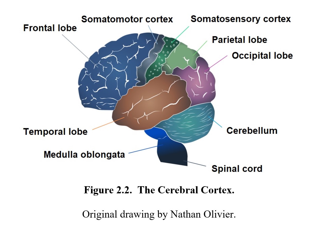
The cortex can be divided into four main lobes. The frontal lobe is situated at the front of the brain and includes a region known as the prefrontal cortex, which is involved in various higher intellectual functions like thought, decision-making, planning, and memory. These skills are referred to as executive function. The frontal lobe also contains the somatomotor cortex or primary motor cortex, which works with other regions of the brain to coordinate skilled voluntary movement. Behind the frontal lobe is the parietal lobe, which integrates sensory information from around the body. It contains the primary somatosensory cortex, the major area for the sense of touch, temperature, and pain. Beneath and lateral to the frontal and parietal lobes is the temporal lobe, which is responsible for auditory processing and some advanced visual processing. It is particularly important for language comprehension. Finally, the occipital lobe resides at the back of the brain and is the visual processing center. It contains the primary visual cortex and allows us to recognize objects and patterns.
The cerebrum (and therefore, the cortex) is divided into right and left hemispheres. Each half controls the opposite side of the body, meaning that a signal to move your right arm comes from the left side of the brain. Though they are essentially symmetrical, the hemispheres are not identical. The left hemisphere is associated with speech and analytical thought, while the right side is associated with creativity, abstract thought, and emotion. While the two hemispheres appear to have their own domains, it does not mean that they are absolutely separate.
2.2.2. Thalamus, Hypothalamus, and Limbic System
In the interior of the brain, hidden by the cerebral cortex, are various important subcortical regions. These regions are a part of the diencephalon (from diá in Greek for through] and enképhalos for brain), which rests above the midbrain and is the other part of the forebrain. For this class, we will focus on three main regions of it: the thalamus; the hypothalamus; and the limbic system.
The thalamus is a large mass in the center of the brain and is the main relay center of the brain. It connects various parts of the cerebral cortex and other areas and relays information between them like a sort of hub. All sensory information (except olfaction, or smell) passes through the thalamus.
Another important subcortical structure is the hypothalamus (from the Latin hypo for under), which is situated below the thalamus and is an important regulatory center in the brain. Its main function is to maintain homeostasis, which is the stable equilibrium of the internal state of the body. It works with the autonomic nervous system to control body temperature, metabolism, hunger, thirst, and fatigue. The hypothalamus regulates the endocrine system by controlling the secretion of various hormones from the pituitary gland. It also contributes to the limbic system and regulates emotions as well as sexual behavior.
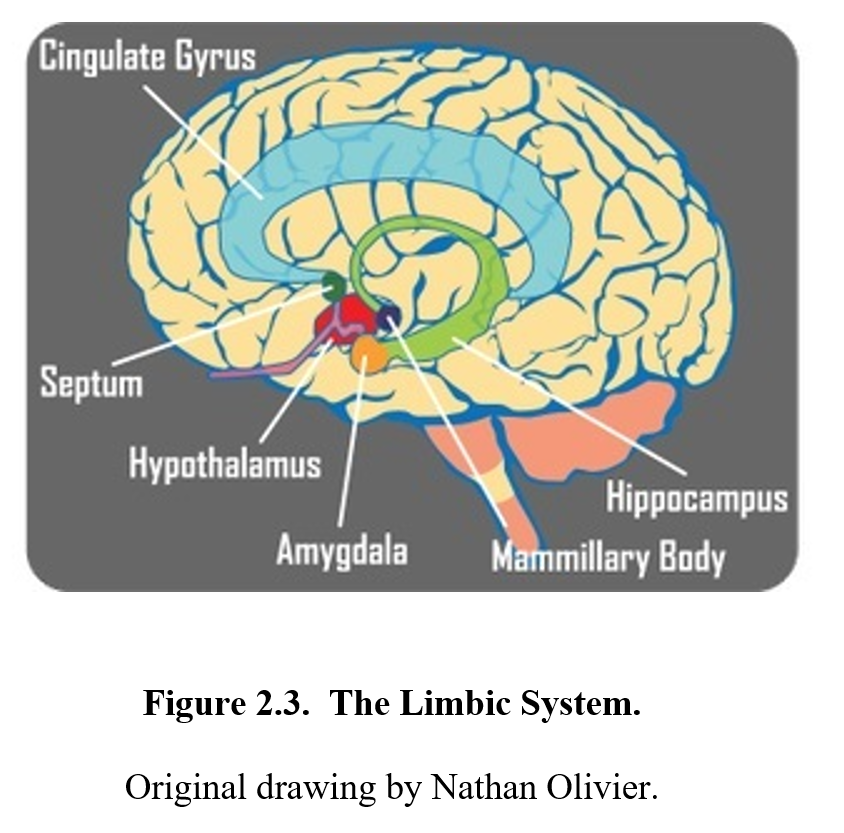
The limbic system encircles the thalamus and is deeply connected to our emotions and motivations, influencing our behavior to ensure our survival. It is not a single discrete structure but an interconnected network of many structures (only a handful of which are relevant to this class). Among these are the amygdala, which is associated with emotional responses such as fear, anxiety, and aggression; the hippocampus, which plays a critical role in storing memories; and the nucleus accumbens, which is an area that is critical in reward, pleasure, and the development of addiction; the cingulate gyrus, which regulates pain and emotion; and the mamillary bodies, which are involved in the retrieval of memories.
2.2.3. Cerebellum and Brainstem
Beneath the forebrain and midbrain is the hindbrain, which consists of the cerebellum and brainstem. The cerebellum (from the Latin for “little brain”) is near the back of the brain, beneath the cerebral cortex, and is smaller and more tightly folded. It is primarily responsible for balance, equilibrium, posture, and coordinating movement. To accomplish this, it receives sensory information from the visual and auditory systems, as well as messages from the cortex and other nerves.
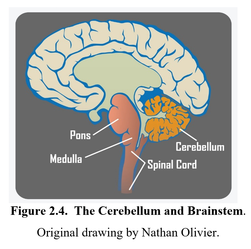
The brainstem joins the spinal cord at the medulla oblongata, which is a regulatory center like the hypothalamus. It oversees breathing, coughing, vomiting, heart rate, blood pressure, and other basic functions. Above the medulla is another brainstem structure called the pons that regulates various functions such as sleep and bladder control and relays signals between the cerebellum and thalamus. A network called the ascending reticular activating system (ARAS) goes through the medulla and pons. It controls the arousal level of the brain, influencing consciousness, alertness, and the sleep/wake cycle.
2.3. Gray Matter vs. White Matter
Section Learning Objectives
- Distinguish gray matter from white matter.
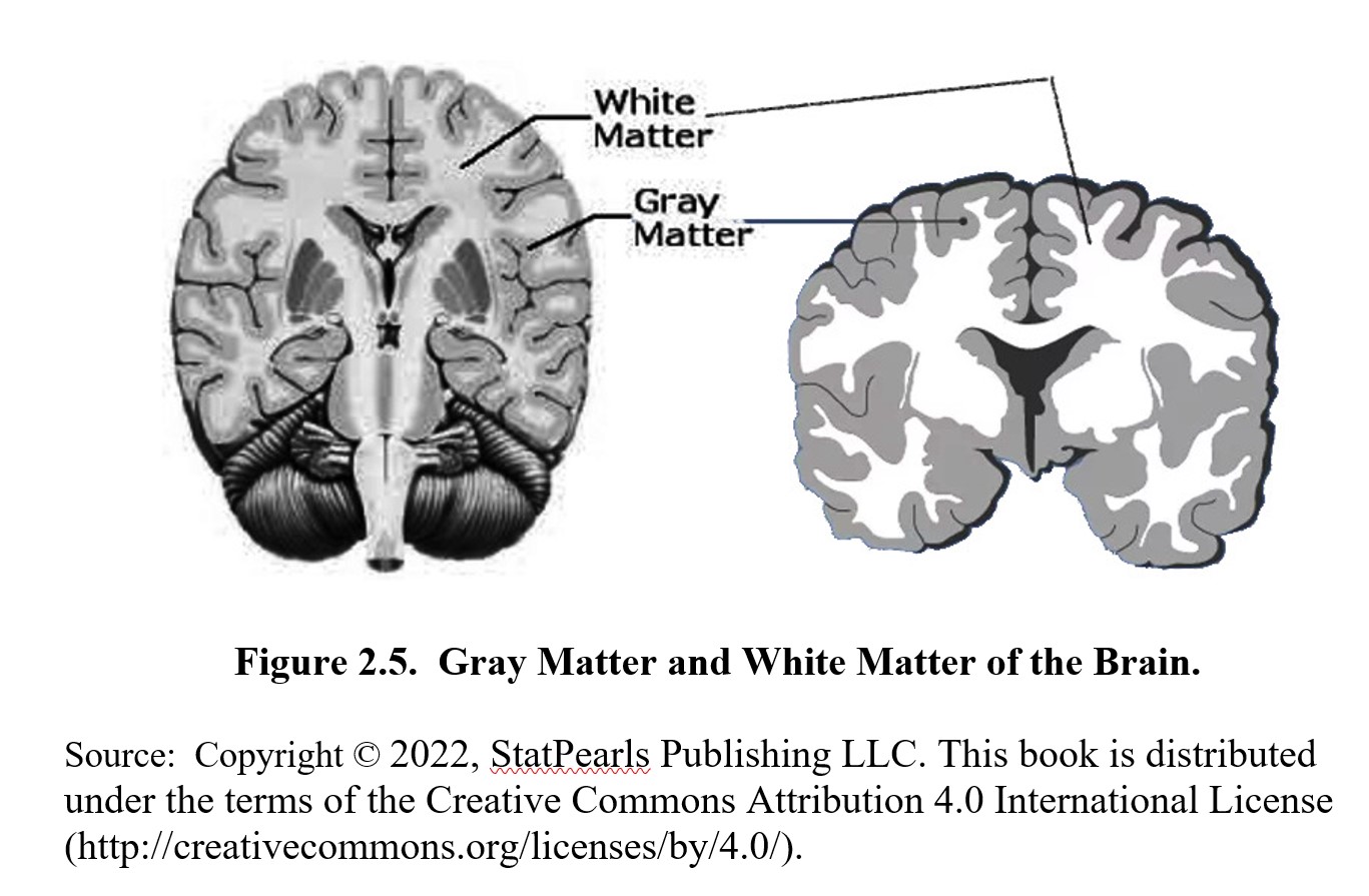
Gray matter is the darker tissue within the brain and spinal cord that is comprised of the neuronal cell bodies, unmyelinated axons, and dendrites as well as glial support cells. Gray matter constitutes 40% of brain tissue. The surface of the cerebral cortex consists of convoluted gray matter divided by gyri and sulci. But located deeper within the cerebral cortex and inside the gyri is white matter, which makes up 60% of brain tissue. It appears whitish in color due to the myelin sheaths that wrap around the nerve fibers. An example of a serious demyelinating neurological disease is multiple sclerosis, abbreviated as MS.
Chapter Recap and Practice Questions
In this chapter, we began our exploration into neuroscience by dividing the nervous system into a central nervous system and a peripheral nervous system. We differentiated between the somatic and autonomic nervous systems and their respective functions. We then took a closer look at the brain in particular and discussed the physiological roles of its major components: the cerebral cortex; thalamus; hypothalamus; limbic system; cerebellum; and brainstem. We will continue our study of the nervous system in the next chapter, so make sure to study the terms in this chapter and check your understanding of the material with the practice questions below before moving on.
Chapter 2 Practice Questions
Answer the following:
- What is the role of the nervous system?
- Explain the difference between a nerve and a neuron.
- What is the difference between afferent and efferent nerve fibers?
- Describe the divisions of the nervous system using six key classification terms. Be sure to explain the function of each division and how each relates to one another.
- What is dual innervation, and why is it important?
- How many lobes are in the cerebral cortex? Give a brief description of the function of each.
- What is the main role of the thalamus in the brain?
- What is the main role of the hypothalamus in the brain?
- Name three structures within the limbic system and define their functions.
- What is the main role of the medulla oblongata?
- Explain the role of the ascending reticular activating system (ARAS)?
- What are the functions of the cerebellum?
2nd edition
