5.5 The Other Senses
Learning Objectives
By the end of this section, you will be able to:
- Describe the basic functions of the chemical senses
- Explain the basic functions of the somatosensory, nociceptive, and thermoceptive sensory systems
- Describe the basic functions of the vestibular, proprioceptive, and kinesthetic sensory systems
Vision and hearing have received an incredible amount of attention from researchers over the years. While there is still much to be learned about how these sensory systems work, we have a much better understanding of them than of our other sensory modalities. In this section, we will explore our chemical senses (taste and smell) and our body senses (touch, temperature, pain, balance, and body position).
THE CHEMICAL SENSES
Taste (gustation) and smell (olfaction) are called chemical senses because both have sensory receptors that respond to molecules in the food we eat or in the air we breathe as opposed to vision that transduces light, and audition that transduces sound waves. There is a pronounced interaction between our chemical senses of taste and smell indicating they work together to provide additional information for estimating the contents of something based on its taste or smell. For example, when we describe the flavor of a given food, we are really referring to both gustatory and olfactory properties of the food working in combination. The senses of taste and smell are able to detect an incredible amount of differences between chemical compounds that inform us about the availability of food, and potential danger or pleasure that could be involved in consuming these compounds. Smell and taste also can initiate and cue gustation and digestion properties, and can also be used as cues for social interactions as in the case of some animals detecting pheromones that cue innate behavioral or physiological responses. The human nose has roughly 400 different types of chemo receptors that allow us to detect at least one trillion different types of odors (Bushdid, Magnasco, Vosshall & Keller, 2014). There are differences between species however where some features of chemosensation have been preserved through evolution and adaptations exist that allow species to utilize chemosensation most effectively in their specific environment.
Taste (Gustation)
You have learned since elementary school that there are four basic groupings of taste: sweet, salty, sour, and bitter. Research demonstrates, however, that we have at least six taste groupings. Umami is our fifth taste. Umami is actually a Japanese word that roughly translates to yummy, and it is associated with a taste for monosodium glutamate (Kinnamon & Vandenbeuch, 2009). There is also a growing body of experimental evidence suggesting that we possess a taste for the fatty content of a given food suggesting that detection of long chain fatty acids (LCFA) may trigger receptors to elicit physiological changes that affect both food intake and digestive functions (Mizushige, Inoue, & Fushiki, 2007; Besnard, Passilly-Degrace & Khan, 2015).
The sense of taste has evolved to be extremely useful in terms of humans survival through the ages. Our ability to differentiate between so many different tastes drives us away from food or drink that could make us sick and toward food and drink we can use as energy. Were also drawn to salty foods which may contain useful minerals and umami which suggests the presence of proteins essential for cellular maintenance and growth. Molecules from the food and beverages we consume dissolve in our saliva and interact with taste receptors on our tongue and in our mouth and throat. Taste buds are formed by groupings of taste receptor cells with hair-like extensions that protrude into the central pore of the taste bud (figure below). Taste buds have a life cycle of ten days to two weeks, so even destroying some by burning your tongue won’t have any long-term effect; they just grow right back. Taste molecules bind to receptors on this extension and cause chemical changes within the sensory cell that result in neural impulses being transmitted to the brain via different nerves, depending on where the receptor is located. Taste information is transmitted to the medulla, thalamus, and limbic system, and to the gustatory cortex, which is tucked underneath the overlap between the frontal and temporal lobes (Maffei, Haley, & Fontanini, 2012; Roper, 2013).

(a) Taste buds are composed of a number of individual taste receptors cells that transmit information to nerves. (b) This micrograph shows a close-up view of the tongue’s surface. (credit a: modification of work by Jonas Töle; credit b: scale-bar data from Matt Russell)
In accordance with the nutritional benefits of carbohydrates and proteins, both sweet and umami are the most likely taste categories to create pleasurable sensations in humans and attraction in animals while bitter tastes often found in poisonous plants create an aversive response in animals and humans to prevent injection of toxic substances. Although most of the taste buds in humans are located on the tongue, some are also located on the palate, the pharynx, epiglottis, and upper third of the esophagus. Taste buds also appear in groups called papillae, of which there are three different types of papillae based on the morphology and where they are located. Fungiform papillae are found in the anterior two thirds of the tongue and are peg like structures with the taste bud located at the top. The foliate papillae, located on the posterior edge of the tongue, and the circumvallate papillae, of which there are only in the posterior area of the tongue are surrounded by grooves lined with taste buds. Each fungiform papillae is made up of one to five taste buds, which each circumvallate or foliate papillae contain hundreds of taste buds.
At the base of each taste cell are dendrite branches stemming from axons of the facial nerve (cranial nerve 7), the glossopharyngeal nerve (cranial nerve 9), and the accessory nerve (cranial nerve 10). These nerves transmit the information from the tongue to the nucleus of the solitary tract, a structure in the medulla of the brainstem that stretches vertically upward and acts as a relay point for taste information. From the nucleus of the solitary tract taste information is transmitted to the ventral posterior nucleus of the thalamus. Recall the thalamus is an important structure located right in the middle of the brain that acts as a rely center between information moving up into the brain and to the specific areas of the cortex where that informations is further processed. From the ventral posterior nucleus of the thalamus, taste informations is sent to the gustatory cortex, located within the fold of the anterior temporal lobe in an area known as the anterior insula-frontal operculum as well as to the hypothalamus, a structure that coordinates both the autonomic nervous system as well as activity of the pituitary gland which maintains and changes body temperature, thirst, hunger, and other homeostatic systems related to sleep and emotion.
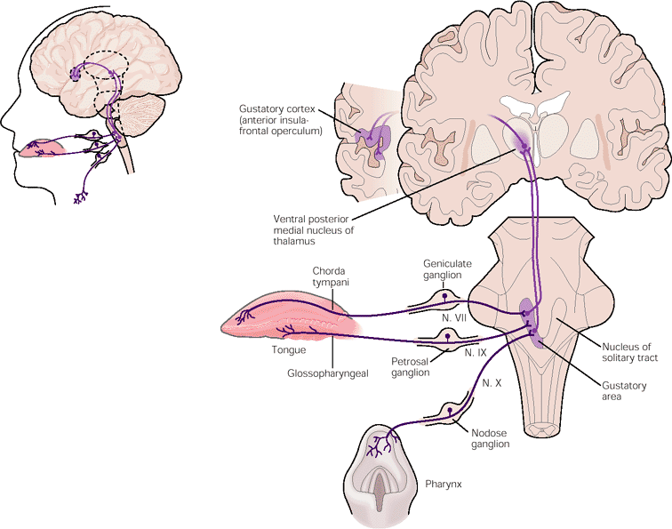 Taste buds transduce chemical information through the tongue and other areas of the mouth to the nucleus of the solitary tract by way of cranial nerves 7, 9 and 10. Information is then relayed to the ventral posterior medial nucleus of the thalamus and the hypothalamus, and finally to the gustatory cortex tucked inside the temporal lobe. Adapted from Hummel, Landis & Hüttenbrink, 2011).
The gustatory cortex is thought to create the conscious perception and discrimination between various tastes. Recordings of electrical activity from the gustatory cortex have suggested that some neurons respond to multiple classes of tastes whereas some respond to only one taste type such as bitter, or sweet. Some believe that the coding of individual tastes may be more related to innate responses attraction to sweet tastes or avoiding something that probably contains poison whereas other groups of neurons encode blends of tastes for sensations. In addition to all the information transduced from by the tastebuds on various parts of the tongue, much of what we know and understand about the flavor of something is also provided from smells transmitted through the olfactory system.
Smell (Olfaction)
Olfactory receptor cells are located in a mucous membrane known as the olfactory epithelium at the top of the nose inside the nasal cavity. The olfactory sensory neuron is a bipolar neuron that extends from the apical end to the epithelial surface, there is branches out with numerous thin cilia that exist within the mucus that coats the nasal cavity. Cilia, Small hair-like extensions from at the ends of these bipolar cells serve as the sites for odor molecules dissolved in the mucus to interact with chemical receptors located on these extensions (figure below). Once an odor molecule has bound to a given receptor, chemical changes within the cell result in signals being sent to the olfactory bulb: a bulb-like structure at the tip of the frontal lobe where the olfactory nerves begin. From the olfactory bulb, information is sent to regions of the limbic system and to the primary olfactory cortex, which is located very near the gustatory cortex (Lodovichi & Belluscio, 2012; Spors et al., 2013) Odorant receptors are created by way of a multigene family that can be found in all vertebrate species (Kandel, Schwartz, Jessel, Siegelbaum & Hudspeth, 2013).

Olfactory receptors are the hair-like parts that extend from the olfactory bulb into the mucous membrane of the nasal cavity.
Olfactory information as figure 3 indicates is transmitted through the olfactory epithelium in the nasal cavity to the olfactory bulb through the bipolar sensory neurons. The olfactory bulb then relays the signals to various areas of the brain such as the anterior olfactory nucleus and the periform cortex located in the interior of the temporal lobe, the amygdala and hypothalamus related to emotion and body autonomic regulation respectively, and the entorhinal cortex and hippocampus related to the maintenance and storage of memories.
There is tremendous variation in the sensitivity of the olfactory systems of different species. We often think of dogs as having far superior olfactory systems than our own, and indeed, dogs can do some remarkable things with their noses. There is some evidence to suggest that dogs can “smell” dangerous drops in blood glucose levels as well as cancerous tumors (Wells, 2010). Dogs’ extraordinary olfactory abilities may be due to the increased number of functional genes for olfactory receptors (between 800 and 1200), compared to the fewer than 400 observed in humans and other primates (Niimura & Nei, 2007).
Many species respond to chemical messages, known as pheromones, sent by another individual (Wysocki & Preti, 2004). Pheromonal communication often involves providing information about the reproductive status of a potential mate. So, for example, when a female rat is ready to mate, she secretes pheromonal signals that draw attention from nearby male rats. Pheromonal activation is actually an important component in eliciting sexual behavior in the male rat (Furlow, 1996, 2012; Purvis & Haynes, 1972; Sachs, 1997). There has also been a good deal of research (and controversy) about pheromones in humans (Comfort, 1971; Russell, 1976; Wolfgang-Kimball, 1992; Weller, 1998).
TOUCH, THERMOCEPTION, AND NOCICEPTION
A number of receptors are distributed throughout the skin to respond to various touch-related stimuli (figure below). These receptors include Meissner’s corpuscles, Pacinian corpuscles, Merkel’s disks, and Ruffini corpuscles. Meissner’s corpuscles respond to pressure and lower frequency vibrations, and Pacinian corpuscles detect transient pressure and higher frequency vibrations. Merkel’s disks respond to light pressure, while Ruffini corpuscles detect stretch (Abraira & Ginty, 2013).

There are many types of sensory receptors located in the skin, each attuned to specific touch-related stimuli.
In addition to the receptors located in the skin, there are also a number of free nerve endings that serve sensory functions. These nerve endings respond to a variety of different types of touch-related stimuli and serve as sensory receptors for both thermoception (temperature perception) and nociception (a signal indicating potential harm and maybe pain) (Garland, 2012; Petho & Reeh, 2012; Spray, 1986). Sensory information collected from the receptors and free nerve endings travels up the spinal cord and is transmitted to regions of the medulla, thalamus, and ultimately to somatosensory cortex, which is located in the postcentral gyrus of the parietal lobe.
Pain Perception
Pain is an unpleasant experience that involves both physical and psychological components. Feeling pain is quite adaptive because it makes us aware of an injury, and it motivates us to remove ourselves from the cause of that injury. In addition, pain also makes us less likely to suffer additional injury because we will be gentler with our injured body parts. Pain perception is a subjective process meaning that it is something that is only accurately understood by the individual experience the pain and that the pain experience can be different for different people who are experiencing the same injury. Many wounded soldiers for example, report not feeling pain until they are actually removed from the battlefield. Injured athletes have reported not being aware of pain related to an injury they suffered until the game or match is over. These examples provide evidence that pain is not the reaction to specific sensory event but is created through the contributions from many different sensory processes and neural signals.
Many periphery organs (organs outside the spinal cord, brainstem, and brain of the central nervous system) such as the skin, joints and muscles contain free nerve endings known as nociceptors that lead to sensory neurons that are able to relay the information to further processing locations. There are three main types of nociceptors that process thermal, mechanical and polymodal (responding to several different forms of sensory stimulation) information and also a fourth class known as silent nociceptors that respond to mechanical stimulation during inflammation and after a tissue injury. Thermal nociceptors nerve endings tend to have a very thin outer layer of myelin fat cells and are activated by extreme differences in temperature, usually greater than 45°C (115°F) and less than 5°C (41°F). Mechanical nociceptors are activated by intense pressure on the skin and are also thinly myelinated. Polymodal nociceptors can be triggered by intense mechanical, chemical or thermal (hot and cold) stimuli and are found at the ends of very small diameter unmyelinated axons that conduct information slower than specifically thermal nociceptors and mechanical nociceptors. Thermal, mechanical and polymodal nociceptors are widely distributed throughout the skin and often activated in larger groups. Silent nociceptors are found in the internal organs in the main cavities of the body such as the abdomen and intestines, and are activated by inflammation and the occurrence of various chemical agents.
Generally speaking, pain can be considered to be neuropathic or inflammatory in nature. Pain that signals some type of tissue damage is known as inflammatory pain. In some situations, pain results from damage to neurons of either the peripheral or central nervous system. As a result, pain signals that are sent to the brain get exaggerated. This type of pain is known as neuropathic pain. Multiple treatment options for pain relief range from relaxation therapy to the use of analgesic medications to deep brain stimulation. The most effective treatment option for a given individual will depend on a number of considerations, including the severity and persistence of the pain and any medical/psychological conditions. Researchers at WSU are studying how much chronic pain can be relieved by long-term opioid pain therapy. Their studies are beginning to show that this form of pain management is not always the most productive: https://news.wsu.edu/2018/07/02/chronic-pain-remains-gets-better-stopping-opioid-treatment/
Persistent pain can be organized into two different categories known as nociceptive pain, and neuropathic pain. Nociceptive pain occurs through the activation of the nociceptors in the skin or soft tissue in response to an injury such as a cut, burn or tissue injury and inflammation. Neuropathic pain on the other hand results from direct damage to nerves in the peripheral or central nervous system and is often reported to the accompanied by burning or electrical sensation (Kandel, Schwartz, Jessel, Siegelbaum & Hudspeth, 2013).
There are five different sensory tracts which transmit pain information from transduction to high level processing in the brain known as the spinothalamic tract, the spinoreticular tract, the spinomesencephalic tract, the cervicothalamic tract, and the spinohypothalamic tract. Signals from the various types of nociceptors in the peripheral nervous system are transmitted from the nerve endings to the cell bodies located in the dorsal root ganglia located and organized in the dorsal section of the spinal cord as vertical layers of perception throughout the body. The spinothalamic tract is the most prominent ascending nociceptive pathway who’s axons cross over the midline of the of the spinal cord at their segment of origin. Signals are then sent to areas of the thalamus and on to the post central gyrus of the cerebral cortex where the the somatosensory cortex is located.
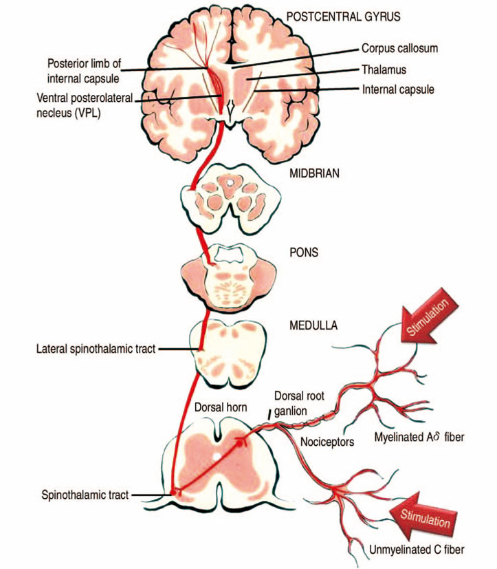 Sensory information is transduced through the open nerve endings of the nociceptors and sent from the cell body in the dorsal horn of the spinal cord, across to the other side of the spinal cord where the signal is sent up to the thalamus by way of the spinothalamic tract. Image adapted from Min and colleagues (2013).
Some individuals are born without the ability to feel pain. This very rare genetic disorder is known as congenital insensitivity to pain (or congenital analgesia). While those with congenital analgesia can detect differences in temperature and pressure, they cannot experience pain. As a result, they often suffer significant injuries. Young children have serious mouth and tongue injuries because they have bitten themselves repeatedly. Not surprisingly, individuals suffering from this disorder have much shorter life expectancies due to their injuries and secondary infections of injured sites (U.S. National Library of Medicine, 2013).
THE VESTIBULAR SENSE, PROPRIOCEPTION, AND KINESTHESIA
The vestibular sense contributes to our ability to maintain balance and body posture. As the figure below shows, the major sensory organs (utricle, saccule, and the three semicircular canals) of this system are located next to the cochlea in the inner ear. The vestibular organs are fluid-filled and have hair cells, similar to the ones found in the auditory system, which respond to movement of the head and gravitational forces. When these hair cells are stimulated, they send signals to the brain via the vestibular nerve. Although we may not be consciously aware of our vestibular system’s sensory information under normal circumstances, its importance is apparent when we experience motion sickness and/or dizziness related to infections of the inner ear (Khan & Chang, 2013).

The major sensory organs of the vestibular system are located next to the cochlea in the inner ear. These include the utricle, saccule, and the three semicircular canals (posterior, superior, and horizontal).
In addition to maintaining balance, the vestibular system collects information critical for controlling movement and the reflexes that move various parts of our bodies to compensate for changes in body position. Therefore, both proprioception (perception of body position) and kinesthesia (perception of the body’s movement through space) interact with information provided by the vestibular system.
These sensory systems also gather information from receptors that respond to stretch and tension in muscles, joints, skin, and tendons (Lackner & DiZio, 2005; Proske, 2006; Proske & Gandevia, 2012). Proprioceptive and kinesthetic information travels to the brain via the spinal column. Several cortical regions in addition to the cerebellum receive information from and send information to the sensory organs of the proprioceptive and kinesthetic systems.
SUMMARY
Taste (gustation) and smell (olfaction) are chemical senses that employ receptors on the tongue and in the nose that bind directly with taste and odor molecules in order to transmit information to the brain for processing. Our ability to perceive touch, temperature, and pain is mediated by a number of receptors and free nerve endings that are distributed throughout the skin and various tissues of the body. The vestibular sense helps us maintain a sense of balance through the response of hair cells in the utricle, saccule, and semi-circular canals that respond to changes in head position and gravity. Our proprioceptive and kinesthetic systems provide information about body position and body movement through receptors that detect stretch and tension in the muscles, joints, tendons, and skin of the body.
References:
Openstax Psychology text by Kathryn Dumper, William Jenkins, Arlene Lacombe, Marilyn Lovett and Marion Perlmutter licensed under CC BY v4.0. https://openstax.org/details/books/psychology
Exercises
Review Questions:
1. Chemical messages often sent between two members of a species to communicate something about reproductive status are called ________.
a. hormones
b. pheromones
c. Merkel’s disks
d. Meissner’s corpuscles
2. Which taste is associated with monosodium glutamate?
a. sweet
b. bitter
c. umami
d. sour
3. ________ serve as sensory receptors for temperature and pain stimuli.
a. free nerve endings
b. Pacinian corpuscles
c. Ruffini corpuscles
d. Meissner’s corpuscles
4. Which of the following is involved in maintaining balance and body posture?
a. auditory nerve
b. nociceptors
c. olfactory bulb
d. vestibular system
Critical Thinking Question:
1. Many people experience nausea while traveling in a car, plane, or boat. How might you explain this as a function of sensory interaction?
2. If you heard someone say that they would do anything not to feel the pain associated with significant injury, how would you respond given what you’ve just read?
3. Do you think women experience pain differently than men? Why do you think this is?
Personal Application Question:
1. As mentioned earlier, a food’s flavor represents an interaction of both gustatory and olfactory information. Think about the last time you were seriously congested due to a cold or the flu. What changes did you notice in the flavors of the foods that you ate during this time?
Glossary:
congenital insensitivity to pain (congenital analgesia)
inflammatory pain
kinesthesia
Meissner’s corpuscle
Merkel’s disk
neuropathic pain
nociception
olfactory bulb
olfactory receptor
Pacinian corpuscle
pheromone
proprioception
Ruffini corpuscle
taste bud
thermoception
umami
vestibular sense
Answers to Exercises
Review Questions:
1. B
2. C
3. A
4. D
Critical Thinking Question:
1. When traveling by car, we often have visual information that suggests that we are in motion while our vestibular sense indicates that we’re not moving (assuming we’re traveling at a relatively constant speed). Normally, these two sensory modalities provide congruent information, but the discrepancy might lead to confusion and nausea. The converse would be true when traveling by plane or boat.
2. Pain serves important functions that are critical to our survival. As noxious as pain stimuli may be, the experiences of individuals who suffer from congenital insensitivity to pain makes the consequences of a lack of pain all too apparent.
3. Research has shown that women and men do differ in their experience of and tolerance for pain: Women tend to handle pain better than men. Perhaps this is due to women’s labor and childbirth experience. Men tend to be stoic about their pain and do not seek help. Research also shows that gender differences in pain tolerance can vary across cultures.
Glossary:
congenital insensitivity to pain (congenital analgesia): genetic disorder that results in the inability to experience pain
inflammatory pain: signal that some type of tissue damage has occurred
kinesthesia: perception of the body’s movement through space
Meissner’s corpuscle: touch receptor that responds to pressure and lower frequency vibrations
Merkel’s disk: touch receptor that responds to light touch
neuropathic pain: pain from damage to neurons of either the peripheral or central nervous system
nociception: sensory signal indicating potential harm and maybe pain
olfactory bulb: bulb-like structure at the tip of the frontal lobe, where the olfactory nerves begin
olfactory receptor: sensory cell for the olfactory system
Pacinian corpuscle: touch receptor that detects transient pressure and higher frequency vibrations
pheromone: chemical message sent by another individual
proprioception: perception of body position
Ruffini corpuscle: touch receptor that detects stretch
taste bud: grouping of taste receptor cells with hair-like extensions that protrude into the central pore of the taste bud
thermoception
umami: taste for monosodium glutamate
vestibular sense: contributes to our ability to maintain balance and body posture
